Home » Without Label » Chest Muscle Anatomy Diagram / Pectoralis Major Muscle Function Origins Human Anatomy Kenhub Youtube : Anatomy of male muscular system, side view.
Chest Muscle Anatomy Diagram / Pectoralis Major Muscle Function Origins Human Anatomy Kenhub Youtube : Anatomy of male muscular system, side view.
Chest Muscle Anatomy Diagram / Pectoralis Major Muscle Function Origins Human Anatomy Kenhub Youtube : Anatomy of male muscular system, side view.. It consists of seven vertebrae. Be sure to visit the guide for more context and information about muscles of the chest diagram for kids or read some of. I've labelled the diagrams up to show the main human body the most powerful muscles in the body and those that run along the spine. The pectoral region is located on the anterior chest wall. Muscle gross anatomy 12 photos of the muscle gross anatomy gross anatomy of cardiac muscle, gross anatomy of skeletal muscle worksheet, gross muscle anatomy test, muscle gross anatomy quiz, muscular system gross anatomy chapter 10, human muscles, gross anatomy of cardiac muscle, gross anatomy of skeletal muscle worksheet, gross.
The pectoral region is located on the anterior chest wall. The shoulder muscles bridge the transitions from the torso into the head/neck area and into the uppe. It is also the center around which the superior 10 ribs directly or indirectly attached. It also protects several vital organs of the chest, such as the heart, aorta, vena cava, and thymus gland that are located just deep to the sternum. Human muscle system functions diagram facts britannica muscle anatomy for female and male dry erase clipboard two sided muscle
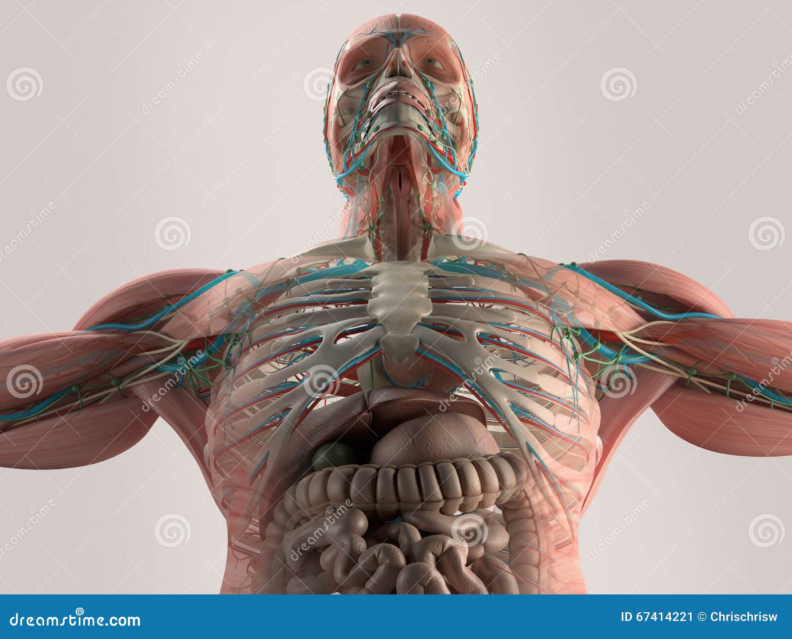
Human Anatomy Chest From Low Angle Bone Structure Veins Muscle On Plain Studio Background Stock Illustration Illustration Of Detail Human 67414221 from thumbs.dreamstime.com The shoulder muscles bridge the transitions from the torso into the head/neck area and into the upper extremities of the arms and hands. The pecs attach to the humerus near the shoulder joint and originate on the breastbone in the center of the chest. See human chest anatomy stock video clips. The neck contains seven of. Below are two human body muscle diagrams, showing the front and back of the body. Hip muscles anatomy hip anatomy anatomy organs human body anatomy human anatomy and physiology anatomy drawing anatomy male shoulder muscle anatomy neck muscle anatomy. Sternocleidomastoid muscle clavicle and ribs anatomy muscle anatomy chest sternocleidomastoid ribs anatomy chest muscles anatomy thorax rib muscles chest muscles chest anatomy illustration. Body_anatomy_muscles_diagram 2/3 body anatomy muscles diagram books body anatomy muscles diagram body anatomy muscles diagram the dominant muscle in the upper chest is the pectoralis major.
Many structures of the chest are readily visible on a chest x ray but others are difficult to see.
It also protects several vital organs of the chest, such as the heart, aorta, vena cava, and thymus gland that are located just deep to the sternum. The pectoralis major, pectoralis minor, serratus anterior and subclavius. Of the two chest muscles, the pectoralis major (a.k.a. Chest anatomy muscles human, chest muscle anatomy diagram, female chest muscle anatomy diagram, human muscles, chest anatomy muscles human, chest muscle anatomy diagram, female chest muscle anatomy diagram. Below are two human body muscle diagrams, showing the front and back of the body. The chest anatomy includes the pectoralis major pectoralis minor and the serratus anterior. Sternocleidomastoid muscle clavicle and ribs anatomy muscle anatomy chest sternocleidomastoid ribs anatomy chest muscles anatomy thorax rib muscles chest muscles chest anatomy illustration. Anatomy of the chest muscles. The pectoral region is located on the anterior chest wall. The sternum is located along the body's midline in the anterior thoracic region just deep to the skin. It also protects several vital organs of the chest, such as the heart, aorta, vena cava, and thymus gland that are located just deep to the sternum. It is also the center around which the superior 10 ribs directly or indirectly attached. Several muscles that move the arms, head, and neck have their origins on the sternum.
Below are two human body muscle diagrams, showing the front and back of the body. The neck contains seven of. Anatomy of the head and upper neck a quick lesson to help you learn more about your craniovertebral junction condition. Skeletal, cardiovascular, nervous and lymphatic systems. Human muscle system functions diagram facts britannica muscle anatomy for female and male dry erase clipboard two sided muscle
Chest Muscles Diagram Quizlet from o.quizlet.com It also protects several vital organs of the chest, such as the heart, aorta, vena cava, and thymus gland that are located just deep to the sternum. Chest muscles diagram 12 photos of the chest muscles diagram anatomy of the chest muscles diagram. Anatomy of male muscular system, side view. Man head and chest anatomy diagram with ghost effect. The neck contains seven of. Be sure to visit the guide for more context and information about muscles of the chest diagram for kids or read some of. 12 photos of the chest muscle anatomy diagram. (as my anatomy professor used to say, if you learn nothing about chest anatomy, learn that the lungs are not in the mediastinum.
This e anatomy module presents an illustrated anatomy of the lungs trachea bronchi pleural cavity and pulmonary vessels.
Skeletal, cardiovascular, nervous and lymphatic systems. Human muscle system functions diagram facts britannica muscle anatomy for female and male dry erase clipboard two sided muscle Below are two human body muscle diagrams, showing the front and back of the body. The sternum is located along the body's midline in the anterior thoracic region just deep to the skin. The pectoral region is located on the anterior chest wall. Chest muscles anatomy for bodybuilders. Body_anatomy_muscles_diagram 2/3 body anatomy muscles diagram books body anatomy muscles diagram body anatomy muscles diagram the dominant muscle in the upper chest is the pectoralis major. The neck contains seven of. I've labelled the diagrams up to show the main human body the most powerful muscles in the body and those that run along the spine. (as my anatomy professor used to say, if you learn nothing about chest anatomy, learn that the lungs are not in the mediastinum. Your chest muscles are big and can handle more weight, which allows you to burn. Diagram of the mediastinum (image source.) in the diagram. The interpretation of a chest film requires the understanding of basic principles.
This e anatomy module presents an illustrated anatomy of the lungs trachea bronchi pleural cavity and pulmonary vessels. Plus, how to target each to make them bigger and stronger. 12 photos of the chest muscle anatomy diagram. Below is a diagram showing the chest muscles depicting where the different exercises target. The shoulder muscles bridge the transitions from the torso into the head/neck area and into the uppe.
Muscle Anatomy Skeletal Muscles Groin Muscles Calf Muscles from healthjade.net An anatomy lesson is a good place to start. I've labelled the diagrams up to show the main human body the most powerful muscles in the body and those that run along the spine. This is a table of skeletal muscles of the human anatomy. Hip muscles anatomy hip anatomy anatomy organs human body anatomy human anatomy and physiology anatomy drawing anatomy male shoulder muscle anatomy neck muscle anatomy. Man head and chest anatomy diagram with ghost effect. The pectoralis major, pectoralis minor, serratus anterior and subclavius. The fibers of the pectoralis muscles run like a fan across the chest. Your chest muscles are big and can handle more weight, which allows you to burn.
Learn anatomy faster and remember everything you learn.
Start with a pair of dumbbells extended above your chest. It also protects several vital organs of the chest, such as the heart, aorta, vena cava, and thymus gland that are located just deep to the sternum. The chest muscles are made up of the pectoralis major and, underneath that, the pectoralis minor. This diagram depicts chest ray oct.human anatomy diagrams show internal organs, cells, systems, conditions, symptoms and sickness information and/or tips for healthy living. Chest muscles, chest muscle diagram. It consists of seven vertebrae. Several muscles that move the arms, head, and neck have their origins on the sternum. Be sure to visit the guide for more context and information about muscles of the chest diagram for kids or read some of. 08102020 related posts of chest muscles diagram chest muscle anatomy exercises. The neck contains seven of. See human chest anatomy stock video clips. This e anatomy module presents an illustrated anatomy of the lungs trachea bronchi pleural cavity and pulmonary vessels. 12 photos of the chest muscle anatomy diagram.